by Alina Ferguson
Cal Poly Humboldt’s campus has a vertebrate museum, dedicated to skulls, skeletons, skins, and tissue from various mammals and vertebrates, such as rats, mice, and sea mammals native to Humboldt county. This student-run museum has about 15,000 specimens, 9,000 of which are available for viewing.
The museum is now allowing all students a peek into their collections, which include rodents, otters, seals, and taxidermied elk. Students are also able to view the anatomical study processes such as dissection and labeling.
Previously, the collection was only accessible to students within the wildlife major. The students would look through the collection and study the specimens, testing their ability to identify them. Now, for the first time since the museum was established in 1969, the museum is open to all students during their open hours on Monday from 11am to 4pm, and Tuesdays through Fridays 12pm to 5pm.
Shea Daly, a senior who works in the lab and does most of the preparations, extended an open invitation to all students.
“On the last Friday of the month, we are going to have open labs and live dissections, so come in and ask questions,” Daly said. “We are also trying to start up scientific drawing classes.”


Daly added that students from all majors are welcome to come in and watch the process of dissecting and skinning the animals.
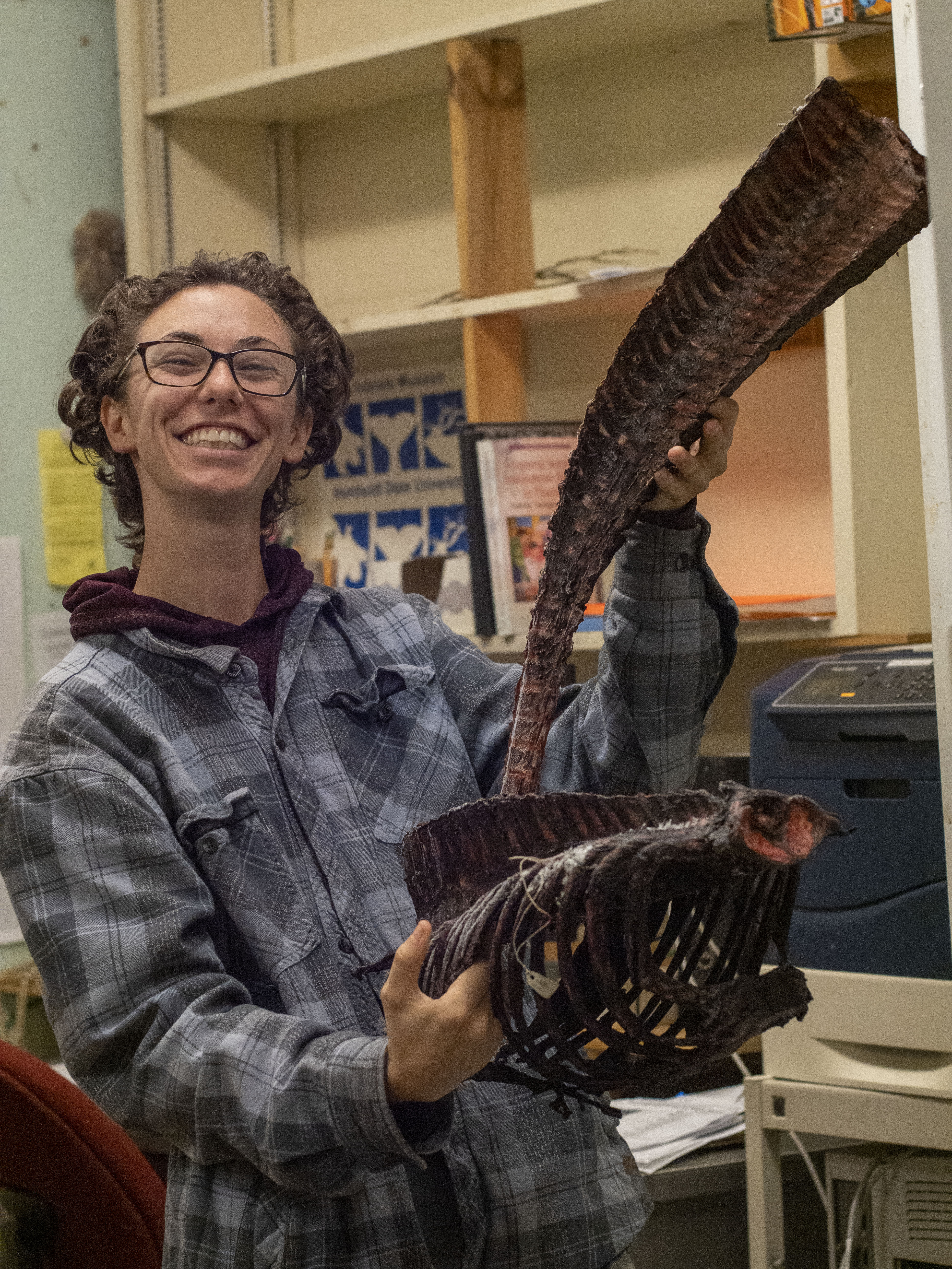
“The students are very hands-on. They can do preps, they can do skeletonizing, which is the process of dipping the bones into an ammonia bath to make them whiter,” Daly said.
In the past, the staff would forage and scrounge for their own specimens. According to Collections Intern Ezra Alberts, now they get most of their specimens from donations, people finding roadkill on the highway, and the marine mammal training program.
Silvia Pavan, the faculty advisor and grant funding organizer to the museum, explained that they typically only need to work with the skull of the animal, as it provides the most information. The rest of the bones are disposed of in a biohazard bin if there is no room for them in the lab or the museum.
Opening a freezer full of numbered vials of tissue samples, Pavan explained the process of tagging each animal and the importance of it. Once an animal has been skinned and deboned, each corresponding part must be labeled and numbered. This tracking lets them trace where the tissue samples came from, the food samples of the animal, and the genealogy of the species.
The museum has a second room, the bug room, where they process the bones that have any remaining flesh or tissue still attached. The bones are placed into a container, where dermestid beetles eat away at everything but bone. Depending on the animal, the carcass may be there for a few days or a few weeks.
A large portion of the work the museum does is tissue sampling, genetics, and CT scanning. This work helps the scientists determine what it ate, where it came from, and ultimately, what killed it.
Leave a Reply